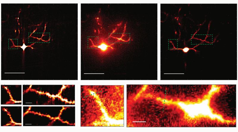
Image courtesy of Yi Xue, et. al.
The human brain, a marvel of nature, is a complex network of neurons that communicate through synapses. Studying these intricate connections is crucial for understanding how our brain functions. However, imaging these synapses in the living brain has been a challenge due to the limitations of existing microscopy techniques. But now, scientists at MIT have developed a new technology that promises to revolutionize this field.
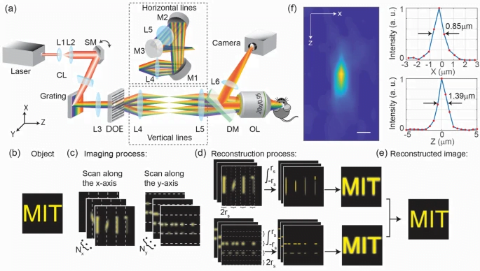
The new technology, called “multiline orthogonal scanning temporal focusing” (mosTF), is a microscopy system designed for fast, clear, and frequent imaging of the living brain. This system works by scanning brain tissue with lines of light in perpendicular directions. Like other live brain imaging systems that rely on two-photon microscopy, this scanning light “excites” photon emission from brain cells that have been engineered to fluoresce when stimulated.
The mosTF system has proven to be eight times faster than a two-photon scope that scans point by point. It also has a four-fold better signal-to-background ratio, a measure of the resulting image clarity, than a two-photon system that just scans in one direction. This significant improvement in speed and clarity is a game-changer for neuroscientists.
The brain’s ability to learn comes from “plasticity,” where neurons constantly edit and remodel the tiny connections called synapses that they make with other neurons to form circuits. To study plasticity, neuroscientists seek to track it at high resolution across whole cells. However, plasticity doesn’t wait for slow microscopes to keep pace, and brain tissue is notorious for scattering light and making images fuzzy.
The mosTF system addresses these challenges effectively. Scanning a whole line of a sample is inherently faster than just scanning one point at a time. While this approach kicks up a lot of scattering, the mosTF system manages that scattering effectively. Some scope systems discard scattered photons as noise, but the mosTF system retains them, improving the clarity of the images.
In conclusion, the mosTF system developed by MIT scientists is a significant advancement in the field of neuroscience. It allows for faster, clearer, and more frequent imaging of the living brain, enabling scientists to study neural circuit connections and plasticity more effectively. This breakthrough could pave the way for new discoveries about how our brain functions and could have far-reaching implications in the study and treatment of neurological disorders.
- Source: MIT